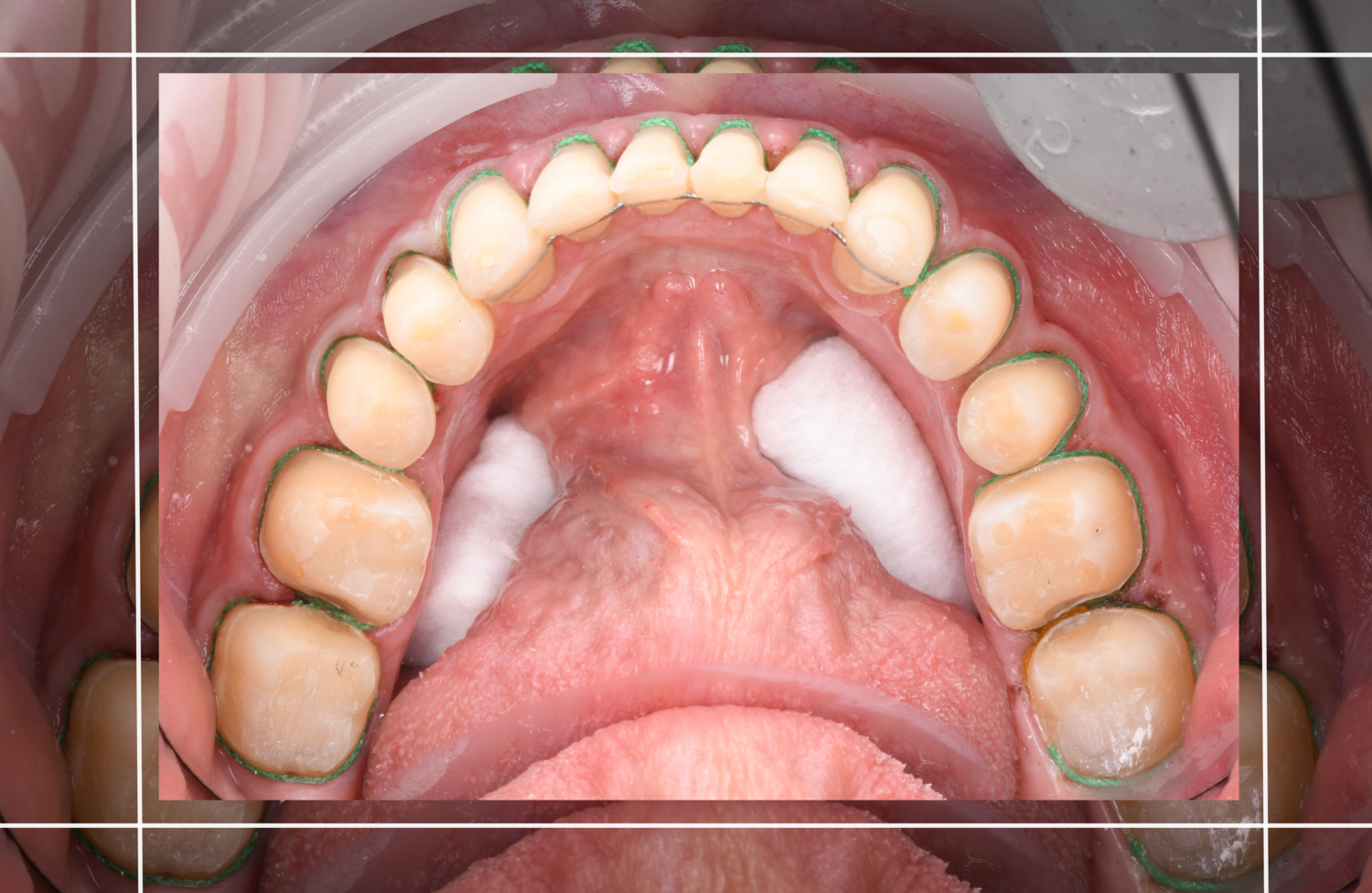
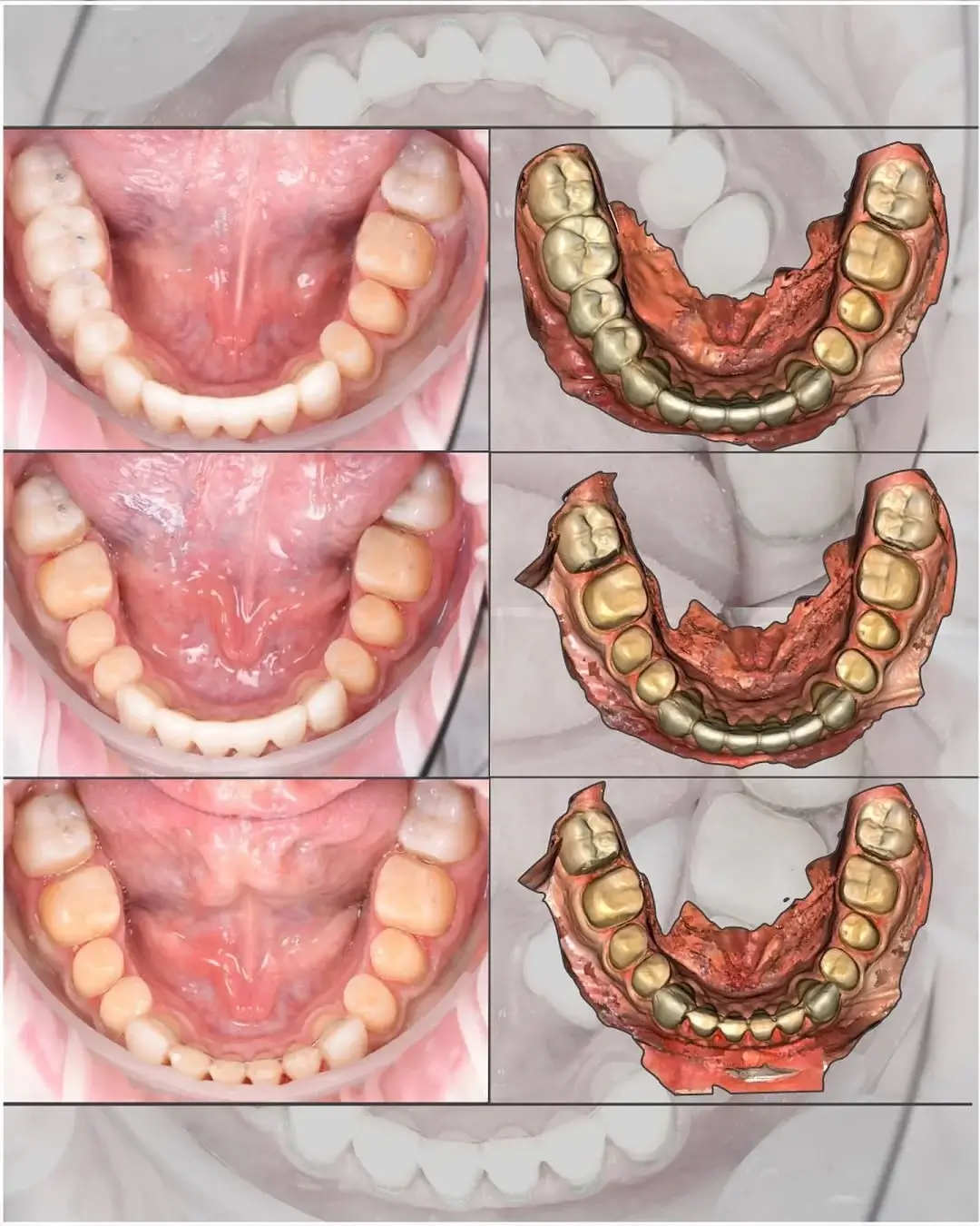
Once we’ve determined the position for reconstruction (CR, new VDO, etc.) and have the models in the articulator in that position where the wax-up is performed, we need to correlate the prep scan with the initial scan (which is already in the articulator). Essentially, the new bite is already created in the articulator.
In this case, I’m demonstrating a situation where the functional mock-up has already been delivered and tested. However, the same process can be done without a mock-up.
Protocol:
1. Initial scan
Perform scans of the upper arch, lower arch, and bite (in centric relation or intercuspal position). If using a mock-up, include it in the scan.

2. Partial tooth preparation
Prepare the posterior teeth, leaving two unprepared (e.g., second molars), and prepare anterior teeth, leaving two unprepared (e.g., canines).
3. Intermediate scan
Capture a scan that includes the four unprepared teeth. These reference points make it easy to align this scan with the 3D models in the digital articulator, where the new mandibular position is pre-established.
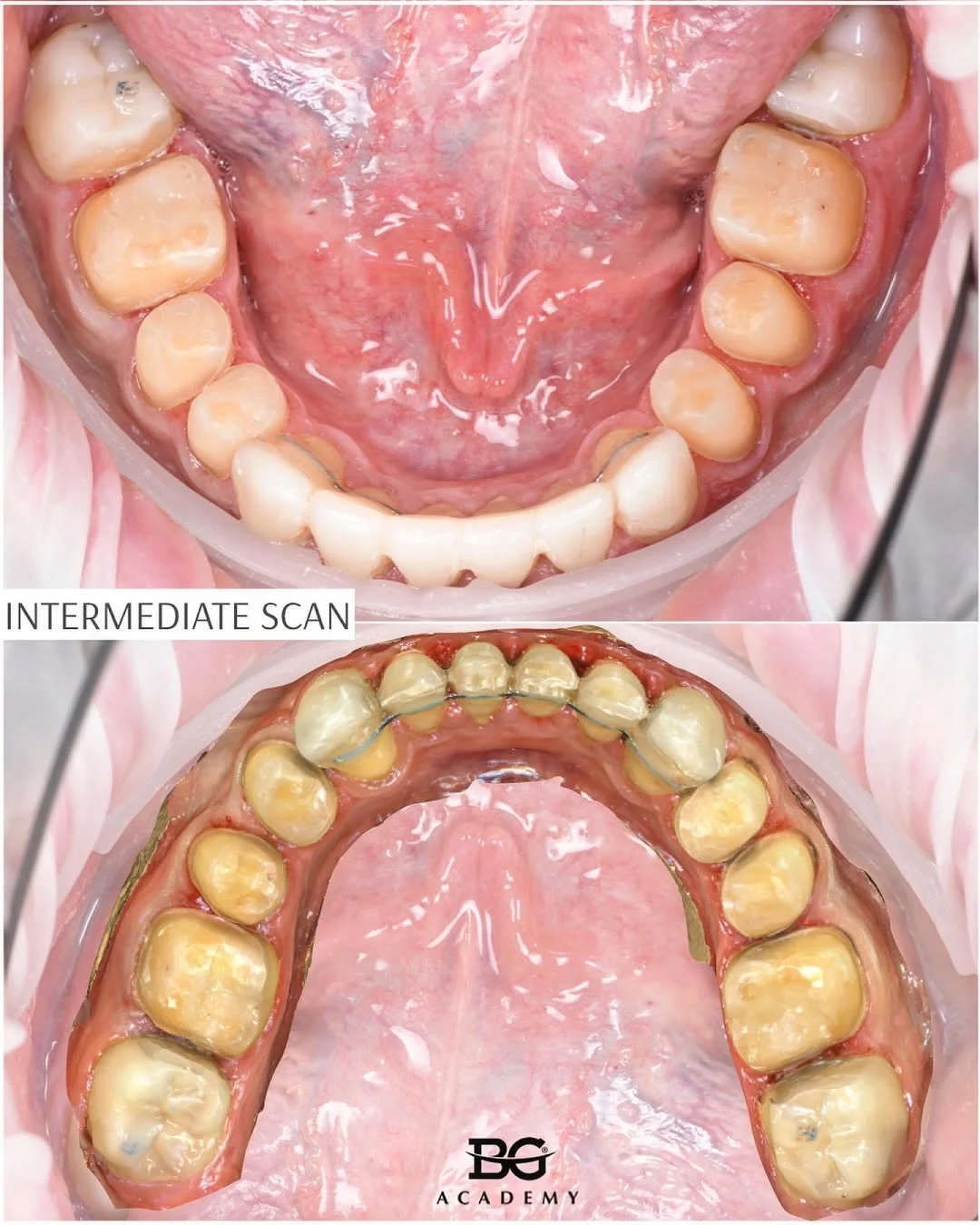
4. Complete tooth preparation and full-arch scan
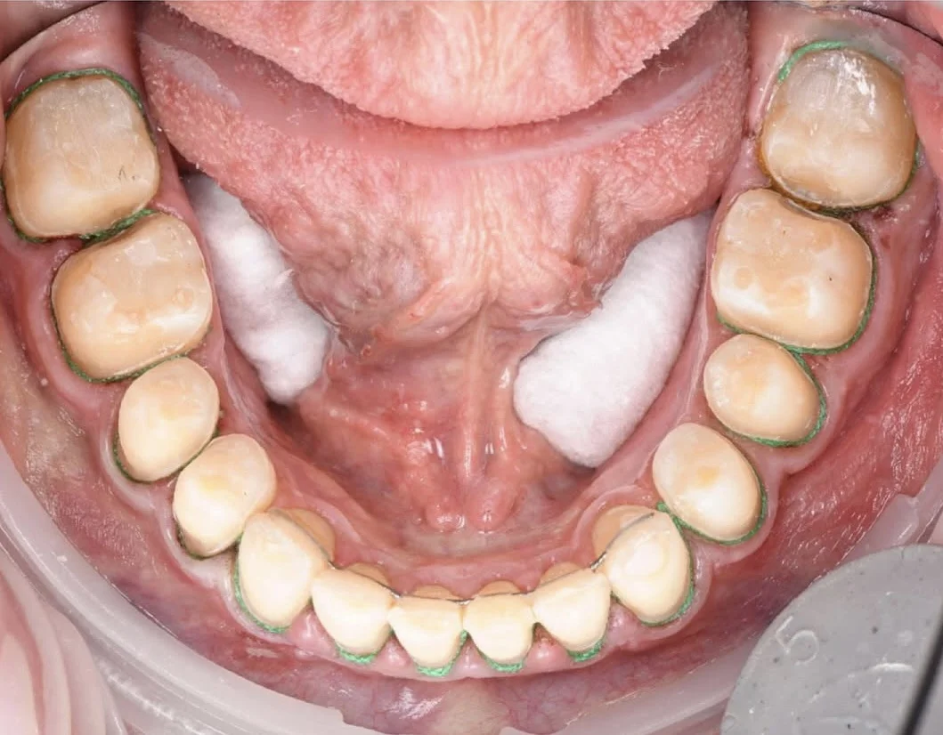
Prepare the remaining teeth (e.g., second molars and canines). Perform a scan of all prepared teeth. This scan is then aligned with the intermediate scan, which is already correlated to the planned bite position in the articulator.
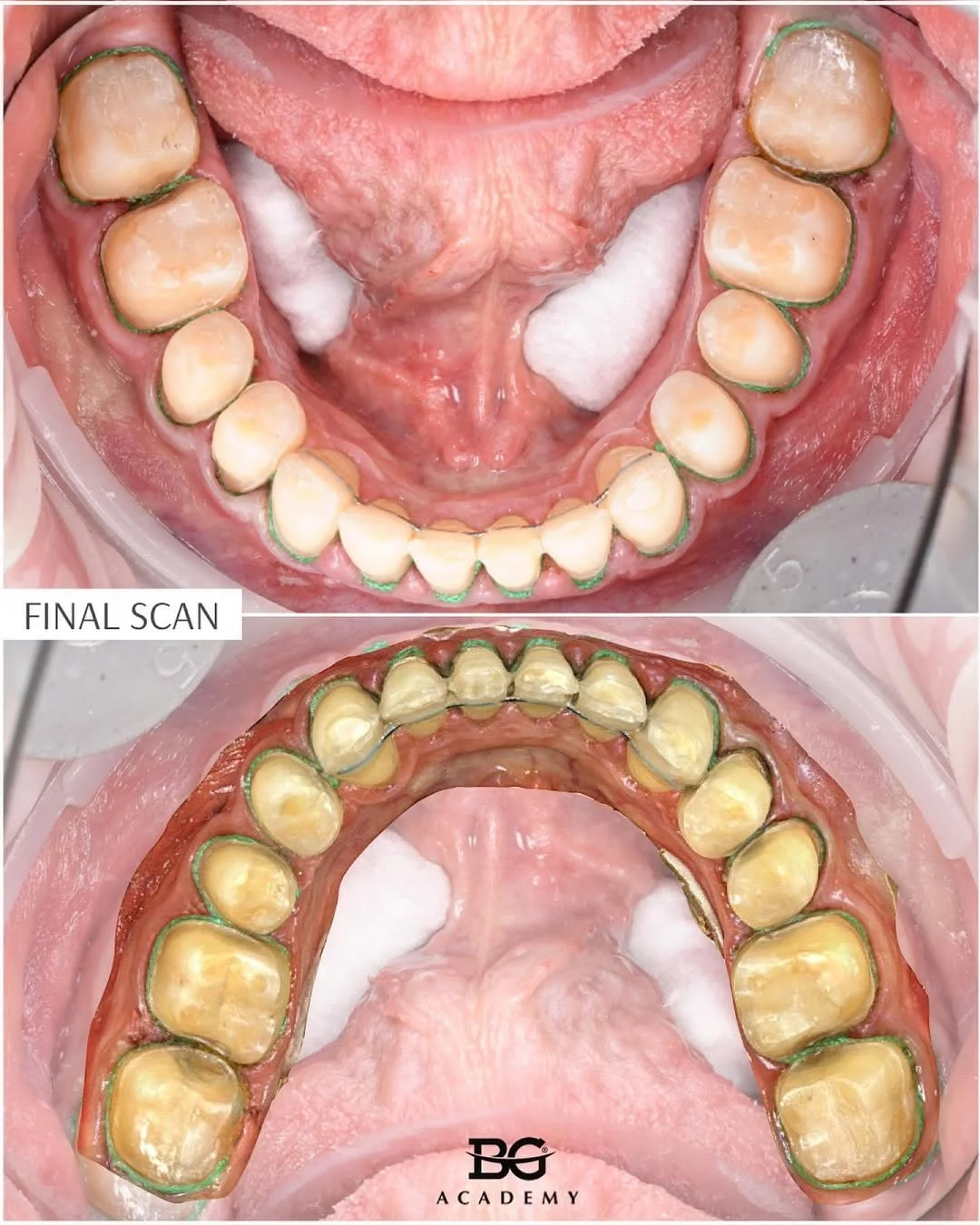
Done.
The most challenging part here is determining and testing the final position in which restorations will be completed. This is part of the treatment planning process that precedes all these preparation steps.
Part of Functional diagnostics and treatment planning course. More info.

